
 Services
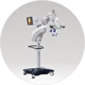
Carl Zeiss Meditec, Germany
Ophthalmic surgical microscope for retinal and glaucoma surgery, cataract treatment, and corneal pathology.
The ZEISS OPMI Lumera 700 is an ophthalmic surgical microscope for retinal and glaucoma surgery, cataract treatment, and corneal pathologies. It is equipped with the integrated ZEISS RESCAN 700 OCT visualization system, which allows visualization of ocular structures in the finest detail and in correct physiological form.
Areas of Application.
Cataract Surgery
The ZEISS OPMI LUMERA 700 provides an unparalleled view of the anterior segment thanks to patented SCI illumination, ZEISS optics, and the ZEISS CALLISTO eye system. The instrument is equipped with an IOL alignment function for efficient and more precise toric IOL alignment with lower residual astigmatism.
The IOL alignment option eliminates the need for manual marking: data is transferred to ZEISS CALLISTO eye from the ZEISS IOLMaster to create overlays in the eyepiece. The ZEISS CALLISTO eye assistance functions are fully controlled by the surgeon via the foot control panel or handle. Time savings and reduced residual astigmatism by eliminating the need for manual preoperative marking, manual data transfer, and manual intraoperative marking. Efficient surgical intervention: real‑time optimization of light intensity, magnification, and image centering with image quality verification. Coaxial illumination provides a stable and high‑contrast red reflex for high detail even in dense cataracts.
Glaucoma Surgery
The ZEISS OPMI LUMERA 700 helps reduce cell loss, shorten manipulation time, and assist the surgeon in making quick decisions during surgery. ZEISS RESCAN 700 in glaucoma surgery and canaloplasty procedures helps monitor stent implants in areas difficult to visualize.
Focus retention: the XY‑axis auto‑tracker maintains focus on the designated area imaged by OCT and compensates for eye or microscope movements. Retinal protection: an integrated retinal protection filter shields the retina from excessive light exposure. Flexibility maintenance: tilt the microscope head as needed to better visualize the iridocorneal angle.
Corneal Pathology Treatment
The integrated intraoperative OCT allows visualization of the physiological corneal shape in two different scanning modes.
Quick switching between high‑resolution OCT scans (scan depth 2.9 mm in tissue) and wide‑field overview images (scan depth 5.8 mm in tissue) for visualization and assessment of graft orientation. Time savings through easy graft monitoring: for DMEK procedures, monitoring graft orientation and interface assessment on the patient’s cornea, as well as checking correct graft positioning. Safe procedure using the Big Bubble technique: improved assessment of dissection depth in deep anterior lamellar keratoplasty using OCT imaging and reduced risk of perforation. The built‑in slit illuminator provides four slit width options with left‑right slit movement for better viewing of the cornea and anterior chamber.
Retinal Surgery
During retinal surgery, the ZEISS OPMI LUMERA 700 microscope provides excellent clarity and a good field of view, as well as an unparalleled view of the anterior segment thanks to patented SCI illumination, ZEISS optics, and the ZEISS CALLISTO eye system.
The built‑in real‑time intraoperative OCT adds a third dimension when viewing transparent ocular media. The XY‑axis auto‑tracker maintains focus on the designated area and compensates for eye/microscope movements. The ability to confirm that all necessary membrane remnants are completely removed after ILM peeling and that there are no macular holes. Detailed retinal images allow surgeons to quickly engage magnification and focus on the area of interest.
Student Training
The ZEISS OPMI LUMERA 700 helps improve the educational process, giving students a clear understanding of the surgical procedure. Whether during surgery, viewing through the assistant’s microscope, or reviewing surgical outcomes, ZEISS provides images with excellent contrast, color, and high resolution.
Enriched experience: ZEISS TRENION 3D HD provides three‑dimensional images that allow students to see what the surgeon sees and gain accurate surgical experience. Recording: use USB devices to document the view through the surgeon’s eyepieces, assistance functions, and intraoperative OCT, with the ZEISS CALLISTO eye system together with a data management system (e.g., ZEISS FORUM) recording the live image to multiple drives. Displaying the surgeon’s eyepiece view allows physicians and students on all connected devices to see what the surgeon sees.