
 Services
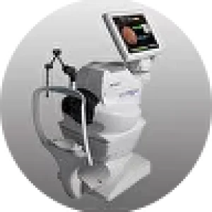
Fully Automated Optical Coherence Tomographer 3D OCT‑1
Topcon, Japan
The fully automated 3D OCT‑1 optical coherence tomographer from Topcon is equipped with a fundus camera and fully automated vascular imaging capabilities. All of this is combined in a single ergonomic device.
3D Wide Scan Width
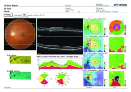
With OCT, a single capture creates a wide‑field 12×9 mm scan. The image covers the macular area and the optic nerve head. This scan is ideal for diagnostics as it significantly reduces examination time. It provides information on retinal thickness and normative data, nerve fiber layers, ganglion cell layers, the GCL+ complex, and topographic information about the optic disc.
True‑Color Fundus Images
The Maestro device incorporates a built‑in fundus camera. With a single capture, OCT and a true‑color fundus photograph are obtained simultaneously. In addition, imaging can be performed with small pupils.
 |  |  |
Follow‑Up Imaging
3D Wide scans provide a full volume of information across a 45° area, but sometimes a scan of smaller areas is needed. In such cases, the Follow‑Up function becomes the optimal choice. Thanks to this, upon repeat visits, the automated tracking program will locate the previously examined area.
Wide Selection of Protocols
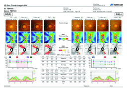
The device offers an extensive range of analytical operations for studying macular pathologies and glaucoma. The resulting reports can be instantly printed or sent to a server.
The Topcon 3D OCT‑1 (Maestro) offers a wide selection of analytical tools for macular pathology diagnostics. Convenient reports can be quickly printed or sent to a server.
Hood Reports
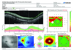
The latest protocols for glaucoma examination can be used with this model. This innovative technology was developed by Dr. Donald Hood. It is based on 3D Wide scans of 12 mm × 9 mm and provides significant…