 Services
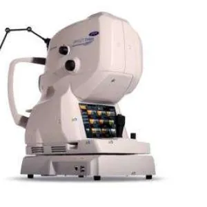
The DRI OCT Triton (Topcon DRI OCT Triton) is a multimodal Swept‑Source optical coherence tomographer (SS‑OCT) from Topcon (Japan). It is one of the first and most renowned swept‑source tomographs on the market, offering extensive capabilities.
Key Specifications
- Technology: Swept‑Source OCT (wavelength 1050 nm).
- Scanning speed: 100,000 A‑scans per second.
- Axial resolution: ~8 µm (digital ~2.6–3 µm).
- Lateral resolution: ~20 µm.
- Penetration depth: Excellent – visualizes from the vitreous body to the sclera and choroid even in the presence of media opacities (cataracts, haemorrhages, gas, etc.).
What the Triton Can Do
- 3D tomography of the posterior segment (macular, disc, wide‑field scans up to 12×9 mm).
- Wide‑field OCT.
- Built‑in fundus camera (true colour non‑mydriatic, red‑free).
- OCTA (angiography without contrast) with OCTARA™ technology.
- Optional: anterior segment (AS‑OCT).
- Triton Plus version adds fluorescein angiography (FA) and autofluorescence (FAF) with Spaide filters.
Key Advantages
- Good penetration through dense cataracts and other opacities (better than many SD‑OCT systems).
- High speed → fewer motion artefacts + eye‑tracking function.
- Multimodality – one device replaces OCT + fundus camera + (in Plus) FA/FAF system.
- Convenient workflow: OCT + fundus photo in one capture, PinPoint™ registration (B‑scan linked to fundus photo).
- PixelSmart™ technology for image quality enhancement.
Primary Clinical Applications
- Diagnosis and monitoring of retinal and macular diseases (AMD, diabetic retinopathy, oedema, epiretinal membranes, etc.).
- Glaucoma (nerve fibre layer analysis, Hood Report).
- Anterior and posterior uveitis, choroidal pathologies.
- Postoperative and post‑injection monitoring.
The Triton is a workhorse for many retinal clinics, valued especially for its reliability in complex patients and its multimodal approach. A newer version, the Triton2, is also available.